Nissl Stains

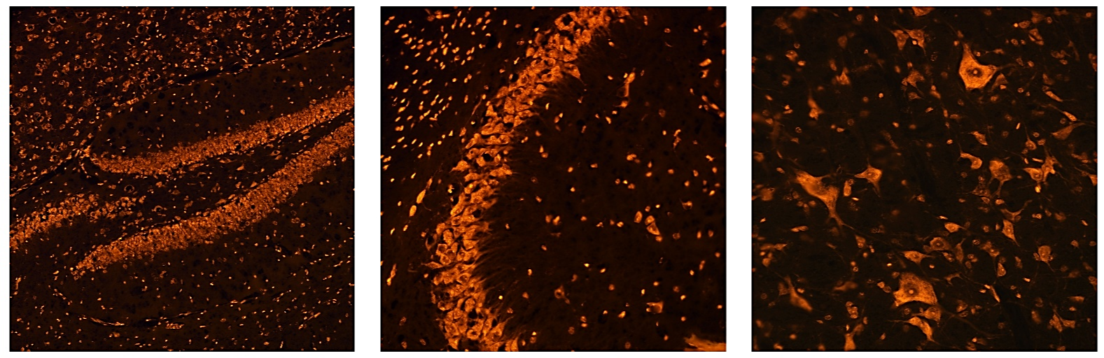
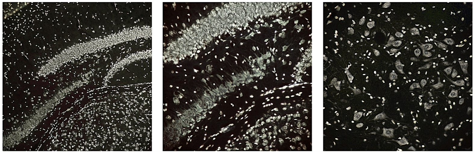
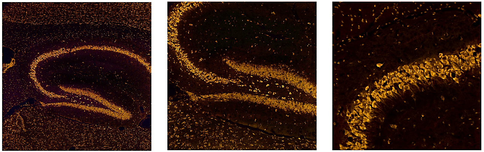
FLUORESCENT NISSL STAINS:
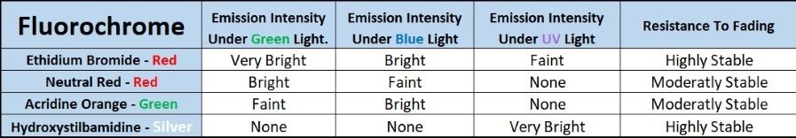
Nissl stains have been a mainstay for studying the neuroanatomy of the brain for over a century by labeling both cellular RNA and nuclear DNA. The increased use of fluorescent microscopy provided a need for fluorescent Nissl stains as well. Therefore, Histo-Chem Inc. is proud to announce 4 different fluorescent Nissl stains provided as a 10X stock solution that can be simply diluted with water 10:1 for histological applications. They may be used in isolation for neuroanatomical studies or in conjunction with another fluorescent tracer for multiple labeling studies. The four fluorescent Nissl stains were selected based on their spectrofluorometric profiles, and are as follows:

New Product 
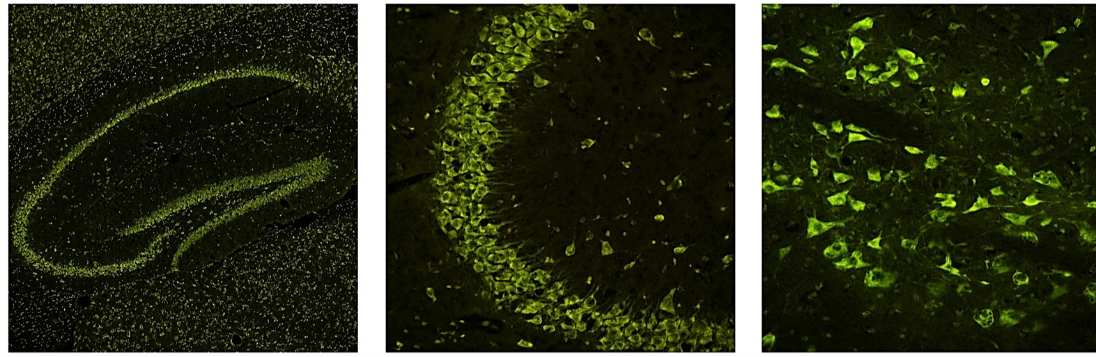
Acridine Orange-Green (#1AOG)
This fluorochrome will stain the cytoplasm and nuclei of neurons a fluorescent green color under blue light excitation. The stain is of high contrast and resolution. The fluorochrome exhibits minimal bleed through when excited by longer or shorter wavelengths and is moderately resistant to fading.

New Product Ethidium Bromide-Red (#1EBR)
This tracer will stain the cytoplasm and nuclei of neurons a fluorescent red color under green light excitation. The stain will be of high contrast, resolution and brightness and will also be very resistant to fading. Although most visible under green light excitation, this fluorochrome will also be moderately visible under blue light excitation and faintly visible under UV excitation.

New Product Hydroxystilbamidine-Silver (#1HS)
This fluorochrome will result in the cytoplasm and nuclei of neurons staining a fluorescent near white color under ultraviolet light excitation. The stain is of very high contrast, brightness and resolution. Also, it will not be visible under longer wavelengths, such as blue or green light excitation. It is also highly resistant to fading.

New Product Neutral Red-Red (#1NRR)
This fluorochrome will also stain the cytoplasm and nuclei of neurons a fluorescent red color under green light excitation. The stain is of relatively high contrast, resolution and is moderately resistant to fading. The fluorochrome shows minimal bleed-through when excited by shorter wavelengths such as blue or UV light.

The below table shows spectrofluorometric properties.

Selecting a Fluorescent Nissl Stain
For routine neuroanatomical studies, the actual color of the tracer is generally of secondary importance. Of primary importance are the brightness, contrast, resolution and permanence of the tracer. For such studies we recommend either the Ethidium Bromide-Red or the Hydroxystilbamadine-Silver. But for multiple labeling studies, we recommend using Neutral Red-Red instead of Ethidium Bromide-Red since the latter will result in noticeable bleed-through under shorter excitation wavelengths. The Acridine Orange-Green can be co localized with tracers excited by either shorter (UV) or longer (green) wavelengths.

Comparing Histo-Chem Inc.’s Fluorescent Nissl Stains with Commercial Cyanine Tracers
Our fluorescent Nissl stains exhibit a number of advantages over fluorescent Nissl stains supplied dissolved in DMSO, such as the NeurotraceTM line. These advantages include greater permanence, brightness, speed and convenience of use. Also, our products are provided in sufficient quantities to be cost-efficient enough to not be limited to on-the-slide staining and therefore can also be used in more convenient Coplin jars or staining dishes. A 4 tracer sample kit (#1NSTP) containing 10 ml of each tracer is also offered. The below table compares the properties of the 2 different fluorescent Nissl stains.

Staining Methodology

  1. Rehydrate tissue sections mounted on gelatin coated slides for 3 min.
  2. Place in the desired fluorescent Nissl staining solution for 10 min. For all 4 tracers the working solution is made by adding 10 ml of stock solution to 90 ml of distilled water.
  3. Rinse slides for 3 min in distilled water.
  4. Differentiate for 3 min in 70% ethanol.
  5. Dehydrate through 2 three min changes of 100% ethanol.
  6. Clear sections with 2 three min changes of xylene.
  7. Mount coverslip with non-polar and non-fluorescent mounting media, such as DPX.