In addition to the manufacture of unique histochemical tracers, Histo-Chem is pleased to offer a brain tissue histology processing and staining service using various unique tracers designed for the localization of specific anatomical structures and pathologies. Brain tissue may be shipped in any aqueous vehicle, such as PBS. Serial or periodic series throughout either a specific brain region or the entire brain are available. Single or multiple brain embedding is also available. Unless otherwise requested, sections are frozen cut at a thickness of 25 um. Sections can then be stained with one or more of Histo-Chem's unique histochemical tracers.
Once the staining is completed, the labeled slides will be shipped in protected slide boxes. Histo-Chem can provide digital imaging of representative slides, if requested. Cost will be based on the number of sections processed and the number of stains used. Please contact us at orders@histo-chem.com for a price quote.
Markers of axonal connectivity:
Fluoro-Gold retrograde axonal transport is used for demonstrating the afferent connectivity of brain regions of interest (1). Examination under UV light following sectioning reveals bright yellow fluorescence of labeled cell bodies and proximal dendrites. Conversely, the anterograde transport of Fluoro-Ruby is used to demonstrate the efferent connectivity of a brain region of interest (2). Examination under green light excitation reveals bright red fluorescent labeling of efferent axons and terminals. In such studies, we can provide the investigator with the appropriate axonal tract tracer for making small intracranial stereotaxic injections. Following suitable survival interval (e.g. 1 -14 days), formalin fixed brains will be dissected and shipped to Histo-Chem for sectioning and staining. Sections will be mounted on gelatin coated slides, dried, stained, cleared and coverslipped with a non-polar non-fading mounting media. Additional in vivo uses for these fluorescent tract tracers include the stereotaxic intraventricular injection of Fluoro-Ruby for the labeling of vascular pericytes (3) and the systemic injection (i.p.) of Fluoro-Gold to label both vascular endothelial cells (4) and CNS neurons with projections to the periphery, including cranial and spinal motor neurons and those with hypothalamic projections to the pituitary.
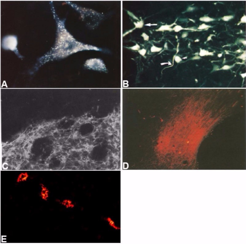
Examples of in vivo staining with Fluoro-Gold or Fluoro-Ruby:
A. Retrogradely labeled neuron within the brainstem reticular formation following Fluoro-Gold injection into the thoracic ventral horn of the spinal cord. B. Neurons within the substantia nigra compacta label with Fluoro-Gold following striatal injection. C. The dorsolateral striatum reveals an extensive network of axons and collaterals following the stereotaxic injection of Fluro-Ruby into the pars reticulata portion of the substantia nigra. D. Dense axons and terminals are seen in the nucleus of the diagonal band following injection of Fluoro-Ruby into the dorsal medial septum. E. Fluoro-Ruby is selectively incorporated into brain vascular pericytes 24 hours following its injection into the lateral ventricle.
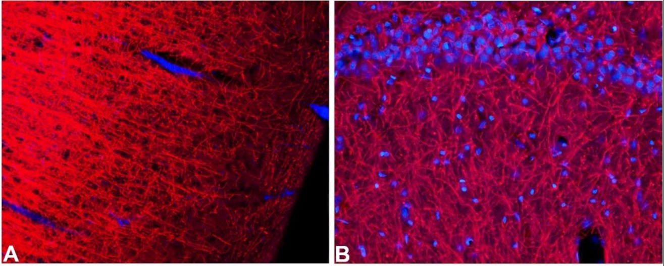
High resolution marker of normal and pathological myelin:
Black-Gold II is a gold based bright-field microscopic stain for the localization of myelin including large tracts as well as fine individual fibers (5). Large myelinated bundles or tracts appear dark brown, while individual fibers appear black in color. The stain can be used to demonstrate either normal myelination or various myelinopathies including edematose swelling, fragmentation and demyelination caused by either neurotoxicant (5) or disease (6). This stain can also be combined with a Nissl stain to simultaneously allow visualization of cell bodies and myelin tracts.
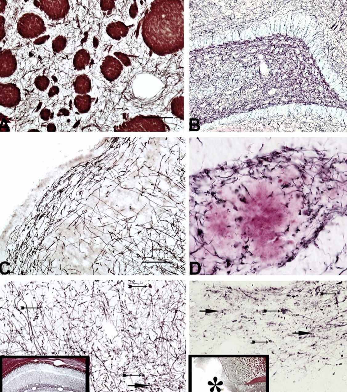
Examples of normal and pathological myelin stained with Black-Gold II:
A. This transverse section through the normal rat striatum exhibits dark brown Black-Gold II staining of large myelinated fascicles as well as the black staining of the interspersed individual fibers. B. Black-Gold II results in detailed staining of individual myelinated fibers in the dentate gyrus region of the rat hippocampus. C. Black-Gold II staining of the piriform cortex reveals both radial myelin in layers II and III as well as fine parallel fibers in layer I. D. Black-Gold II staining of the dentate gyrus of an aged AD/Tg mouse reveals various myelinopathies including fragmentation, edematose swellings and demyelination surrounding a large amyloid plaque counterstained with Congo Red. E. Black-Gold II staining of the rat hippocampus following exposure to kainic acid reveals both edematose and fragmented myelinated fibers. F. Black-Gold II staining of the piriform cortex following exposure to kainic acid reveals regions of frank demyelination (*) as well as fragmentation and swelling of adjacent myelinated fibers.
Localization of degenerating neurons:
Fluoro-Jade B (7) and Fluoro-Jade C (8) are used to localize neurons and their processes that are undergoing irreversible degeneration due to either apoptotic or necrotic insult. Such insults can be due to disease, physical trauma or chemical neurotoxins. Survival times following acute injury typically range from 1-14 days, at which time the formalin fixed brains would be dissected and then shipped to us for sectioning, mounting, staining and coverslipping. We routinely use Fluoro-Jade C, although Fluoro-Jade B is also available for similar staining of degenerating neurons and their processes. Both tracers emit a bright green fluorescence under blue light excitation.
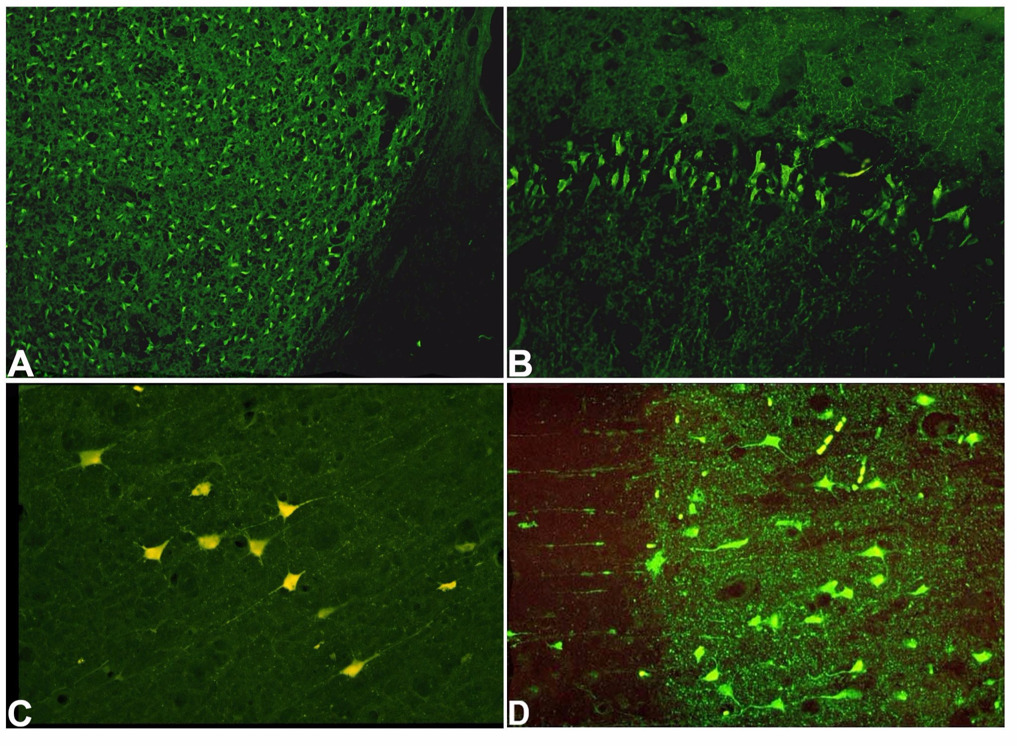
Markers of neuronal degeneration:
A. Extensive Fluoro-Jade C positive degenerating neurons can be seen throughout the striatum following dosing with 3-nitropropionic acid. B. Fluoro-Jade C positive degenerating neurons of the CA1 region of the hippocampus following dosing with methamphetamine. C. Following exposure to MDMA, Fluoro-Jade C positive neurons, proximal dendrites and axonal terminals are seen in the piriform cortex. D. Fluoro-Jade C also stains degenerating neurons, dendrites and axon terminals in the cingulate cortex following exposure to kainic acid.
Localization of amyloid plaques:
Amylo-Glo is used for the localization of amyloid plaques seen in the brains of transgenic animals that model Alzheimer’s disease as well as humans with the disease (9). Under ultraviolet excitation the tracer appears light yellow in color, which typically appears as light blue when photographed. It stains the cores and neurites of various plaques types including dense core, condensed and diffuse morphologies.
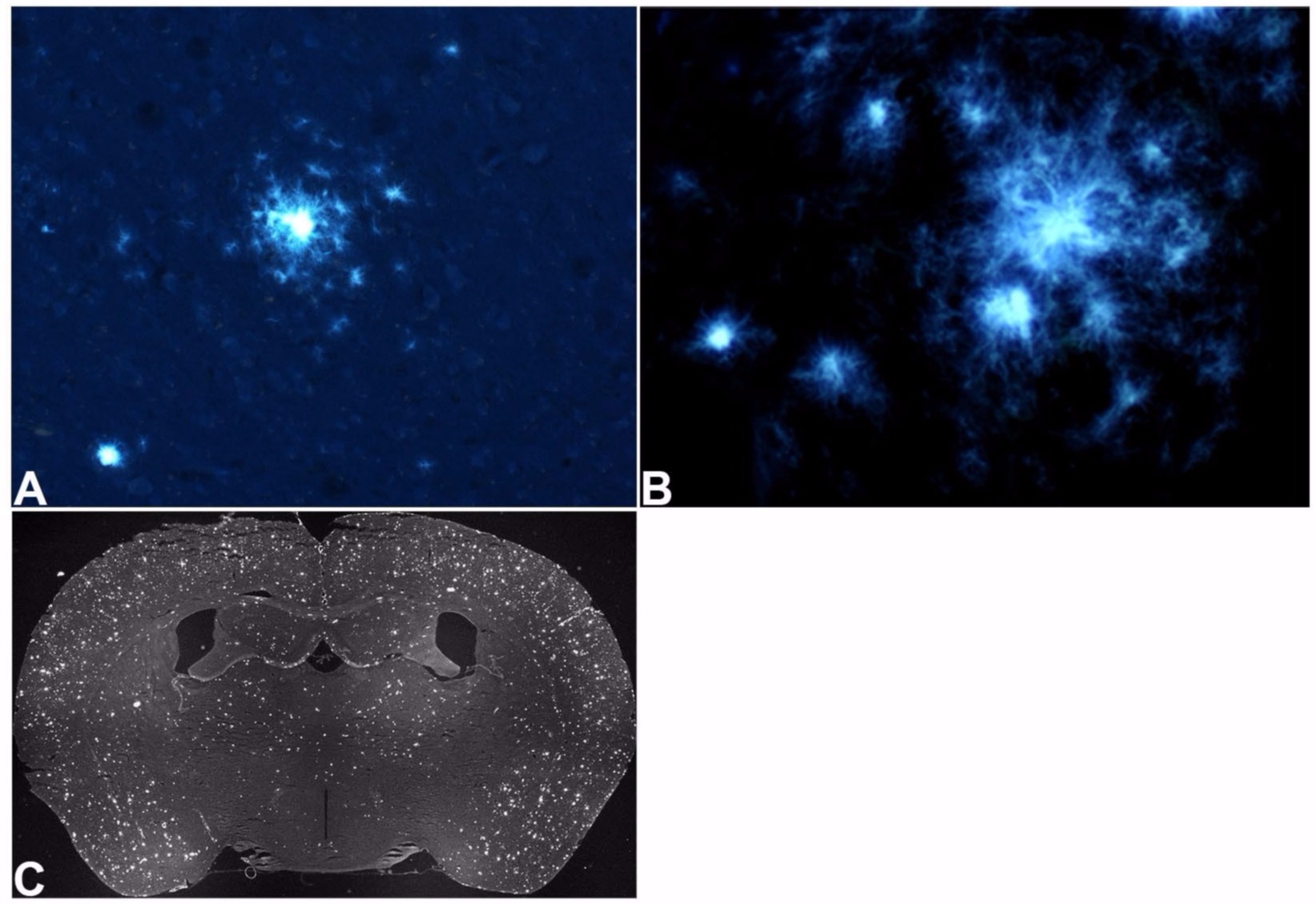
Markers of amyloid plaques:
A. Cortical amyloid plaques from aged AD/Tg mouse labeled with Amylo-Glo. B. High magnification view of amyloid plaques shows both dense core and fibular components. C. Survey view of an entire aged AD/Tg mouse brain section is suitable for both qualitative and quantitative analysis.
Euro-Glo for the localization of myelin and amyloid plaques:
We have developed a different class of tracer that can also be used for localizing amyloid plaques, specifically the fluorescent Europium chelate, Euro-Glo (10). Euro-Glo, under UV excitation, results in a red emission due to its extremely high Stoke’s Shift value. It labels both myelinated fibers and amyloid plaques. This tracer differs from the Amylo-Glo staining in that it is not compatible with the use of nonpolar solvents, requires several days to develop and the amyloid plaques reveals vesicular as well as fibular morphologies. These properties suggest the staining of lipid containing molecules in myelin and plaques.
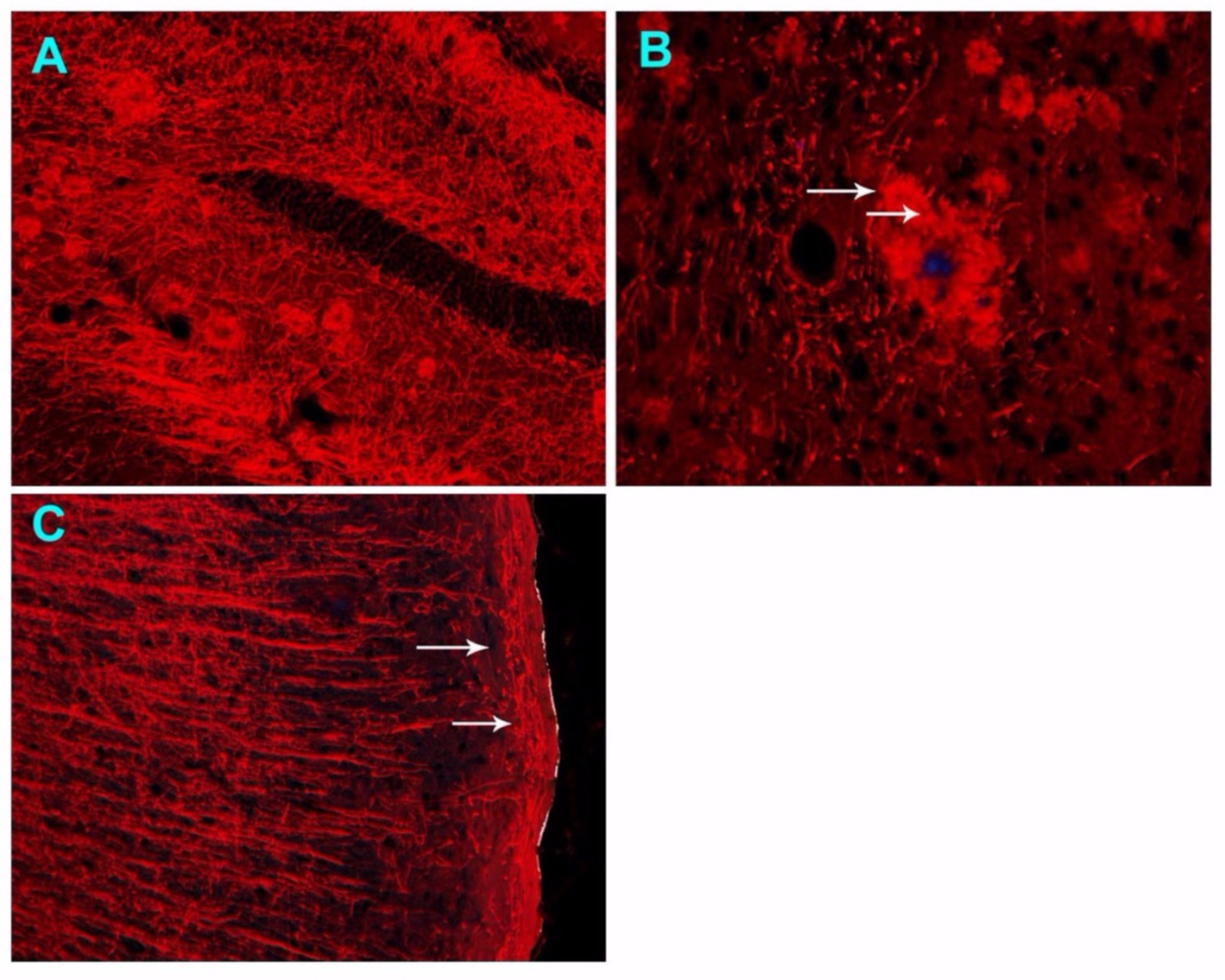
Examples of Euro-Glo labeling:
A. Euro-Glo staining of an aged AD/Tg mouse brain reveals both myelinated fibers as well as amyloid plaques in the hippocampus. B. Higher magnification of Euro-Glo labeled amyloid plaque reveals both a blue colored core and red surrounding fibular and globular appearing structures (arrows). C. Euro-Glo staining of the rat parietal cortex reveals both deep radial and superficial parallel (arrows) myelinated fibers.
Multiple labeling studies:
The combination of more than one histochemical tracer can be very informative and numerous multiple labeling combinations of the aforementioned tracers are possible.
Multiple labeling with fluorescent axonal tract tracers:
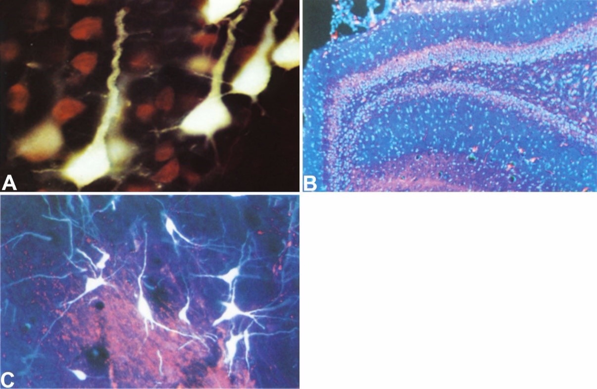
A. Fluoro-Gold labeling of layer V sensory motor neurons with projections to the ventral horn of the spinal cord appear light yellow, while adjacent neurons lacking such projections appear red following counterstaining with ethidium bromide. B. The granule and polymorphic cells of the dentate gyrus appear blue following counterstaining with DAPI while red Fluoro-Ruby transported anterogradely from the dorsomedial septum reveals labeled axon terminals adjacent to these neurons. C. Simultaneous injection of the retrograde axonal tract tracer Fluoro-Gold into the ventral striatum and the anterograde tract tracer Fluoro-Ruby injected into the nucleus accumbens results in Fluoro-Gold labeled cells in the pars compacta and Fluoro-Ruby labeled terminals in the pars reticulata regions of the substantia nigra.
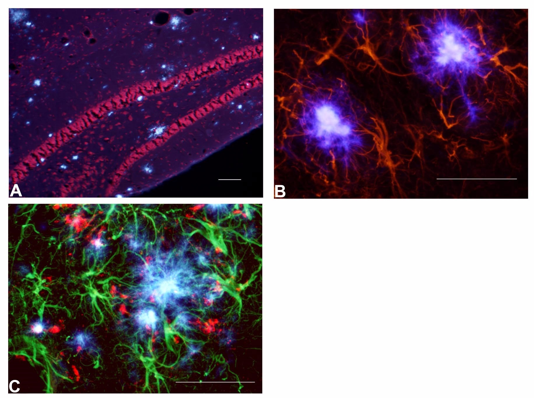
Multiple labeling with Black-Gold II myelin stain:
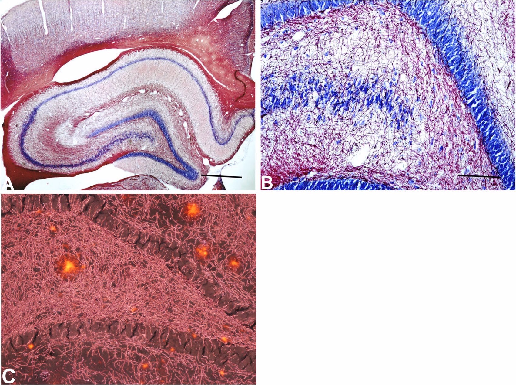
A.-B. Low and high magnification view of the rat hippocampus illustrates the combined Black-Gold II localization of white matter (myelin) and the Toluidine Blue O staining of gray matter (cytoplasmic Nissl substance). C. When viewed under combined fluorescent and darkfield illumination, Black-Gold II labeled fibers are seen in the dentate gyrus except for in the unstained granule cells and the Congo Red fluorescence of amyloid plaques in AD/Tg mice.
Similarly, fluorescent Nissl or nuclear stains may be combined with any of the fluorescent markers previously discussed. For example, counterstaining with ethidium bromide results in the red labeling of cytoplasmic Nissl substance under green light illumination while DAPI staining results in the blue labeling of cellular nuclei when viewed under violet or ultraviolet illumination (11). Also available is double labeling with immunofluorescent markers such as GFAP for the localization of hypertrophied astrocytes (examples below). Inquiries regarding multiple labeling strategies are welcome.
Multiple labeling with fluorescent markers of neuronal degeneration:
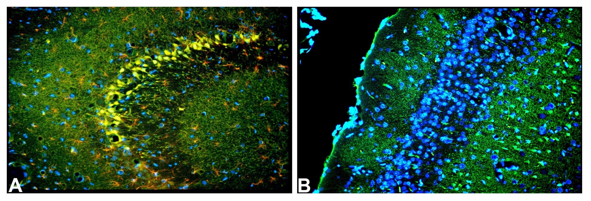
A. The CA1-CA2 region of the hippocampus of a rat given kainic acid used Fluoro-Jade C, anti-GFAP and DAPI for the respective colocalization of degenerating neurons and terminals (green), hypertrophied astrocyte (red) and cell nuclei (blue). B. The cingulate cortex of a rat given kainic acid reveals green Fluoro-Jade C positive degenerating pyramidal neurons in layer III and terminals in layer I, while viable blue DAPI stained granule cells are seen in layer II.
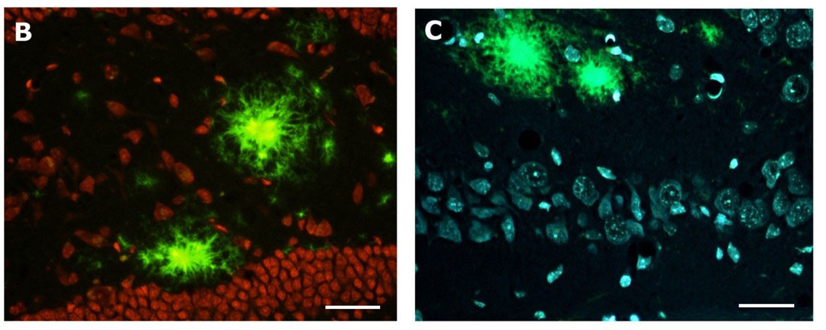
Multiple labeling of Amylo-Glo stained tissue:
A. Blue Amylo-Glo stained plaques are colocalized in the hippocampus of the AD/Tg mouse relative to a red ethidium bromide Nissl counterstain. B.-C. Blue Amylo-Glo labeled amyloid plaques and hypertrophied astrocytes are colocalized with antibodies to GFAP and appear red or green when respectively visualized with TRITC (B) or FITC (C).
Multiple labeling with Euro-Glo:
A. Red Euro-Glo stained myelinated fibers contrast with the lumen of blood vesicles filled by intravascular perfusion with Fluoro-Turquoise labeled gelatin (12) at the time of sacrifice. B. Both gray and white matter in the hippocampus can be labeled by the respective application of Euro-Glo for the red labeling of myelinated fibers and DAPI for the blue labeling of nuclei, including the pyramidal cells of CA1 seen at top.
Multiple labeling with HQ-O:
Incubation of tissue sections from the brain of an AD/Tg mouse model of Alzheimer’s disease in a HQ-O solution results in the bright and specific labeling of amyloid plaques. The labeling has been attributed to zinc bound to the plaques.
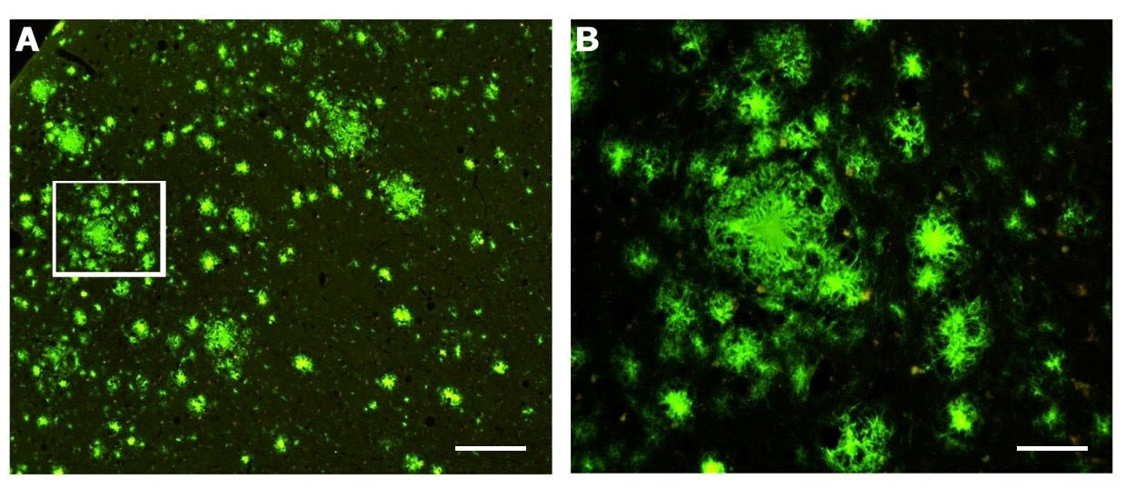
Fig A is a survey view of a section of the cortex of a one year-old APP/PS1 mouse (pial surface at upper left). Box seen at the left is shown in Fig B under higher magnification to show fibular appearance of labeling.
B. Combined HQ-O and Ethidium bromide staining of the dentate gyrus of an AD/Tg mouse reveals the amyloid plaque location relative to other cells in the dentate gurus of the hippocampus, including granule cells seen at bottom. C. Combined HQ-O and DAPI staining of the CA-1 region of the hippocampus reveals the location of amyloid plaques relative to the nuclei of various cells, including the pyramidal neurons seen below the amyloid plaques.
Once the staining is completed, the labeled slides will be shipped in protected slide boxes. Histo-Chem can provide digital imaging of representative slides, if requested. Cost will be based on the number of sections processed and the number of stains used. Please contact us for a price quote.
| REFERENCES | |
|---|---|
| 1 | Schmued, LC and Fallon, JH; Fluoro-Gold: a new fluorescent retrograde axonal tracer with numerous unique properties, Brain Res., 1985, 346(1), 124-9. |
| 2 | Schmued, LC, Kyriakidis, K, Heimer; in vivo anterograde and retrograde axonal transport of the fluorescent rhodamine-dextran-amine, Fluor-Ruby, within the CNS; Brain Res., 1990, 536(1), 127-34. |
| 3 | Sarkar, S and Schmued, LC; In vivo administration of fluorescent dextrans for the specific and sensitive localization of brain vascular pericytes and their characterization in normal and neurotoxin exposed brains; Neurotox., 2012, 33(3), 436-43. |
| 4 | Bowyer, JF, Tranter, KM, Sarkar, S, Raymick, J, Hanig, JP, Schmued, LC; Systemic administration of Fluoro-Gold for the histological assessment of vascular surface, integrity and damage, Curr. Neurovasc. Res., 2014, 11(1), 31-47. |
| 5 | Schmued, LC, Bowyer, JF, Cozart, M, Heard, D, Binienda, Z, Paule, M; Introducing Black-Gold II, a highly soluble gold phosphate complex with several unique advantages for the histochemical localization of myelin; Brain Res. 2008, 1229, 210-7. |
| 6 | Schmued, LC, Raymick J, Paule, MG, Dumas, M Sarkar, S; Characterization of myelin pathology in the hippocampal complex of a transgenic mouse model of Alzheimer’s disease, Curr. Alzheimers Res., 2013, 10(1), 30-7. |
| 7 | Schmued, LC, Hopkins, KJ; Fluoro-Jade B: a high affinity marker for the localization of neuronal degeneration, Brain Res., 2000, 874(2), 123-30. |
| 8 | Schmued, LC, Stowers, CC, Scallet, AC, Xu, L; Fluoro-Jade C results in ultrahigh resolution and contrast labeling of degenerating neurons, Brain Res, 2005, 1035(1), 24-31. |
| 9 | Schmued, LC, Raymick, J, Tolleson, W, Sarkar, S, Zhang, YH, Bell-Cohn, A; Introducing Amylo-Glo, a novel fluorescent amyloid specific histochemical tracer especially suited for multiple labeling and large scale quantification studies, J. Neurosci. Meth., 2012, 209(1), 120-6. |
| 10 | Schmued, LC and Raymick, J; Introducing Euro-Glo, a rare earth metal chelate with numerous applications for the fluorescent localization of myelin and myelin and amyloid plaques in brain tissue sections, J. Neurosci. Meth., 2017, 279, 79-86. |
| 11 | Schmued, LC, Swanson, LW, Sawchenko, PE; Some Fluorescent counterstains for neuroanatomical studies, J. Histochem. and Cytochem., 1982, 30(2),123-8. |
| 12 | Sarkar, S, Raymick, J, Paule, MG, Schmued, LC; In situ demonstration of Fluoro-Turquoise conjugated gelatin for visualizing brain vascular and endothelial cells and their characterization in normal and kainic acid exposed animals, J. Neurosci. Meth., 2013, 219(2), 276-84. |