Multiple Labeling Studies

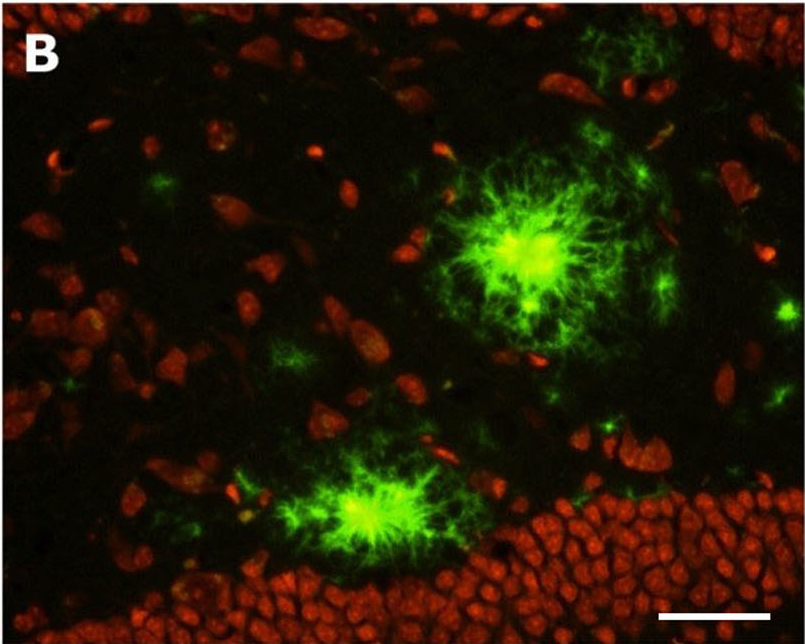
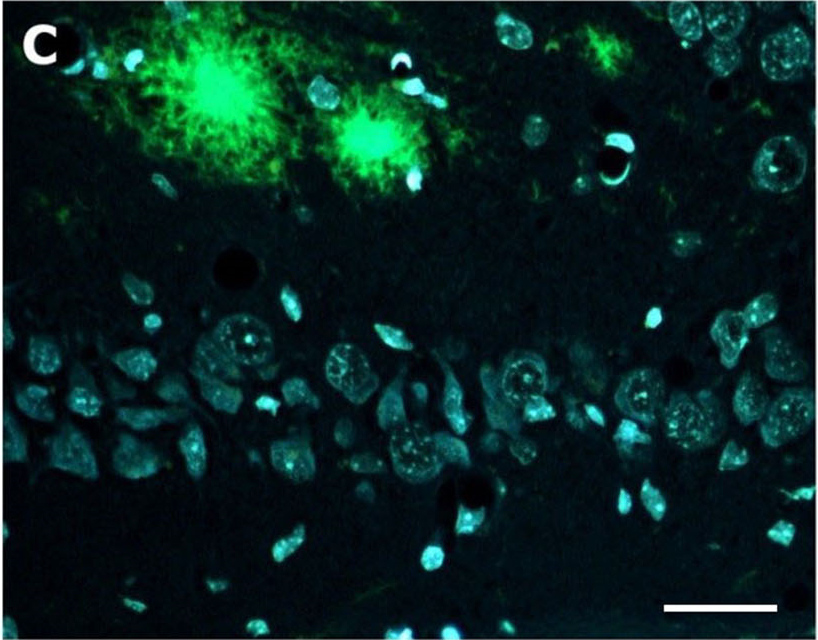
The combination of more than one histochemical tracer can be very informative and numerous multiple labeling combinations of the aforementioned tracers are possible, as the following examples illustrate:

Multiple Labeling with fluorescent axonal tract tracers

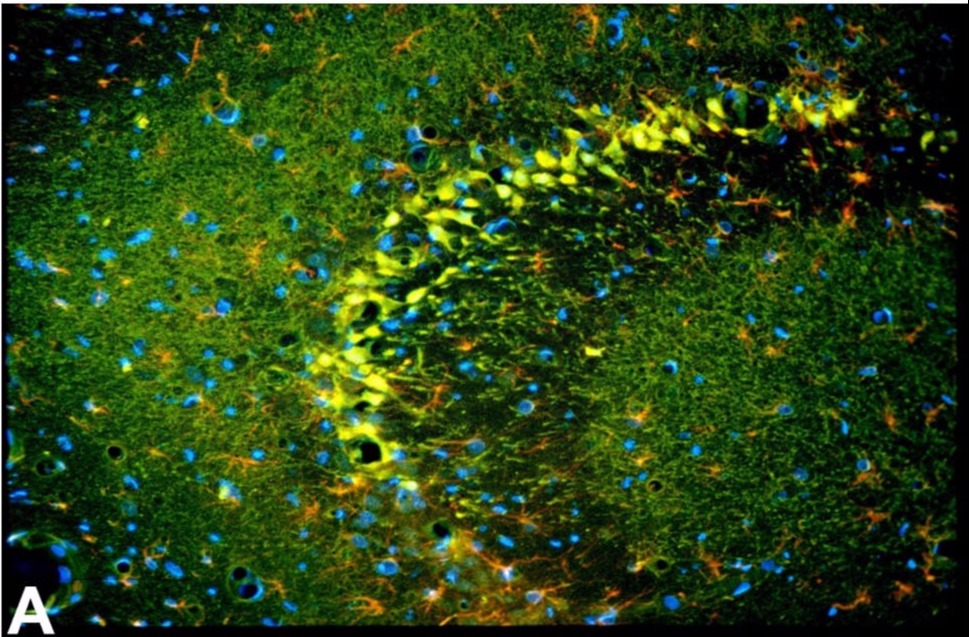
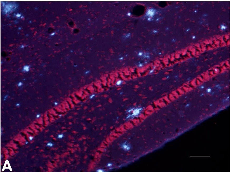
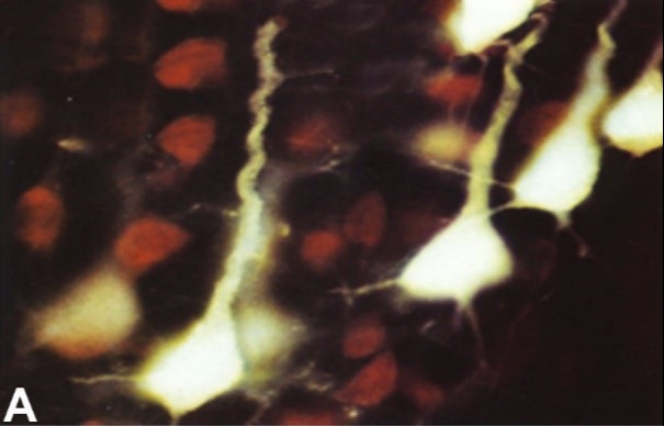
A. Fluoro-Gold labeling of layer V sensory motor neurons with projections to the ventral horn of the spinal cord appear light yellow, while adjacent neurons lacking such projections appear red following counterstaining with ethidium bromide. 

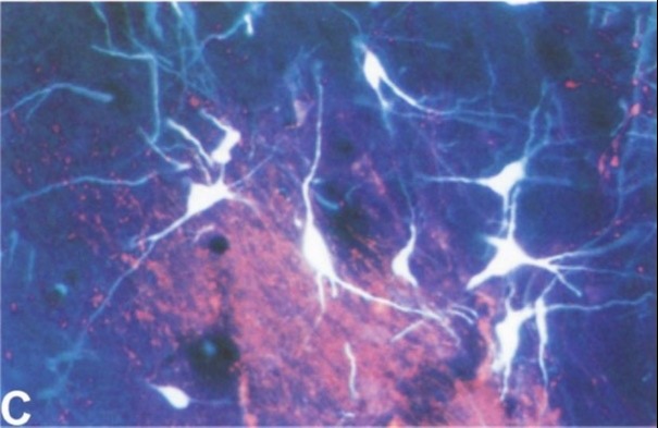
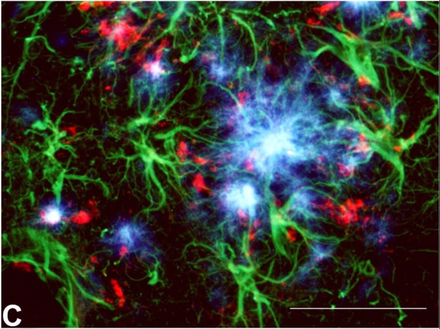
C. Simultaneous injection of the retrograde axonal track tracer Fluoro-Gold into the ventral striatum and the anterograde tract tracer Fluoro-Ruby injected into the nucleus accumbens results in Fluoro-Gold labeled cells in the pars compacta and Fluoro-Ruby labeled terminals in the pars reticulatta regions of the substantia nigra.

Multiple labeling with Black-Gold II myelin stain

A-B. Low and high magnification view of the rat hippocampus illustrates the combined Black-Gold II localization of white matter (myelin) and the Toluidine Blue O staining of gray matter (cytoplasmic Nissl substance).

Similarly, fluorescent Nissl or nuclear stains may be combined with any of the fluorescent markers previously discussed. For example, counterstaining with ethidium bromide results in the red labeling of cytoplasmic Nissl substance under green light illumination while DAPI staining results in the blue labeling of cellular nuclei when viewed under violet or ultraviolet illumination (11). Also feasible is double labeling with immunofluorescent markers such as GFAP for the localization of hypertrophied astrocytes (examples below). Enquiries regarding multiple labeling strategies are welcome.

Multiple labeling with fluorescent markers of neuronal degeneration

A. The CA1-CA2 region of the hippocampus of a rat given kainic acid used Fluoro-Jade C, anti-GFAP and DAPI for the respective colocalization of degenerating neurons and terminals (green), hypertrophied astrocyte (red) and cell nuclei (blue).

B. The cingulate cortex of a rat given kainic acid reveals green Fluoro-Jade C positive degenerating pyramidal neurons in layer III and terminals in layer I, while viable blue DAPI stained granule cells are seen in layer II.

Multiple labeling of Amylo-Glo stained tissue

A. Blue Amylo-Glo stained plaques are colocalized in the hippocampus of the AD/Tg mouse relative to a red ethidium bromide Nissl counterstain.

C. Blue Amylo-Glo labeled amyloid plaques and green hypertrophied astrocytes are colocalized with antibodies to GFAP.

Multiple labeling with Euro-Glo

A. Red Euro-Glo stained myelinated fibers contrast with the lumen of blood vessels filled by intravascular perfusion with Fluoro-Turquoise labeled gelatin (12) at the time of sacrifice.

B. Both gray and white matter in the hippocampus can be labeled by the respective application of Euro-Glo for the red labeling of myelinated fibers and DAPI for the blue labeling of nuclei, including the pyramidal cells of CA1 seen at top.

Multiple labeling with HQ-0

B. Combined HQ-O and Ethidium bromide staining of the dentate gyrus of an AD/Tg mouse reveals the amyloid plaque location relative to other cells in the dentate gurus of the hippocampus, including granule cells seen at bottom.

C. Combined HQ-O and DAPI staining of the CA-1 region of the hippocampus reveals the location of amyloid plaques relative to the nuclei of various cells, including the pyramidal neurons seen below the amyloid plaques.