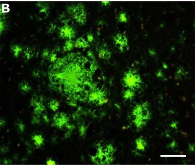
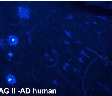
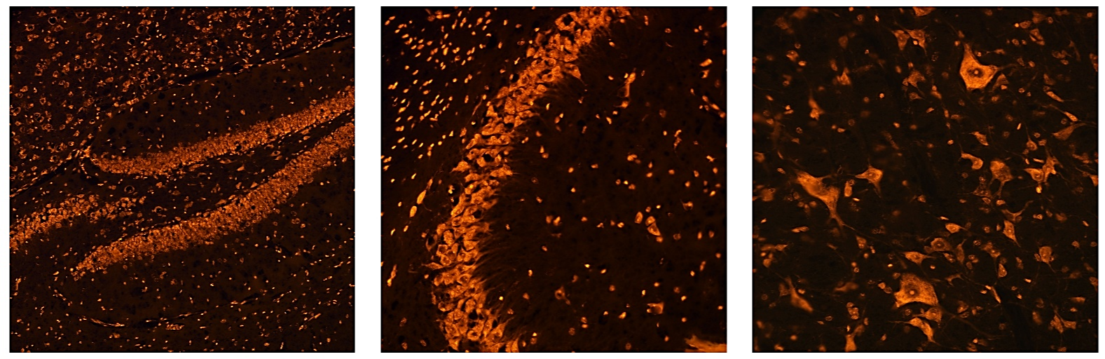
New & Future Dyes

Recently Developed

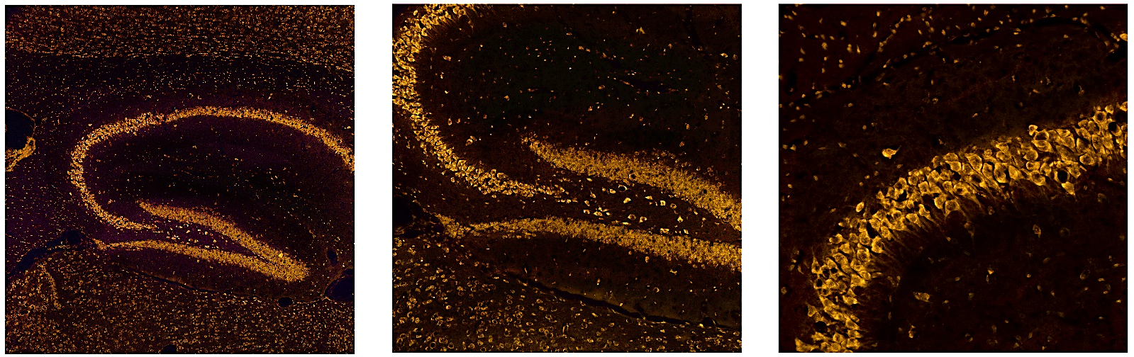
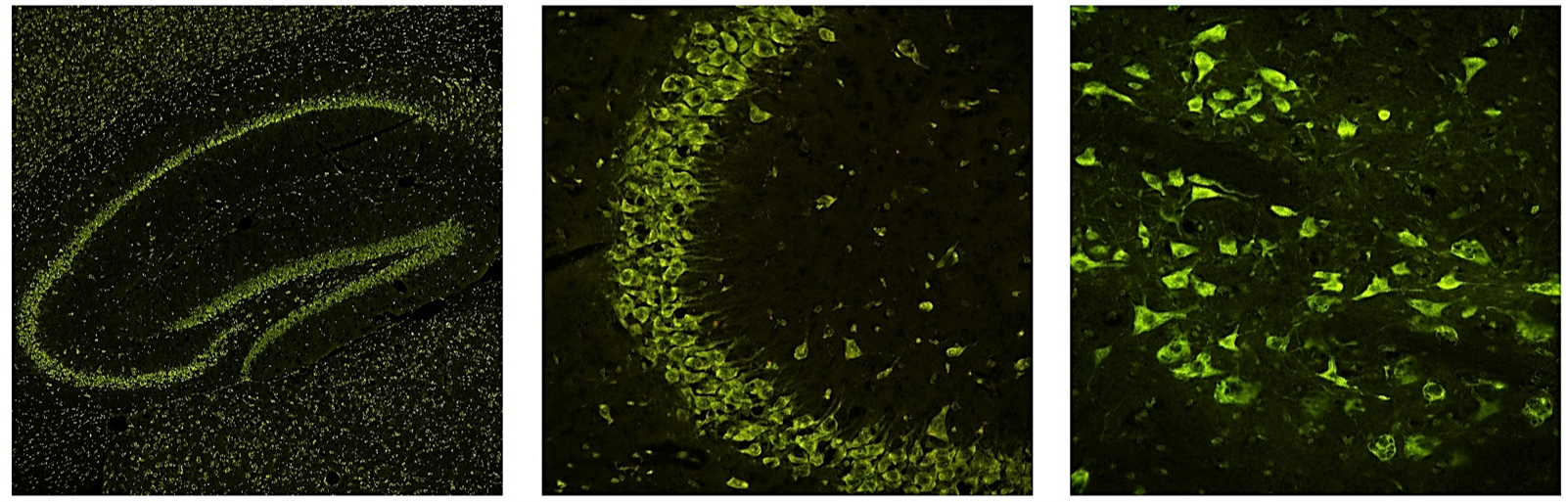
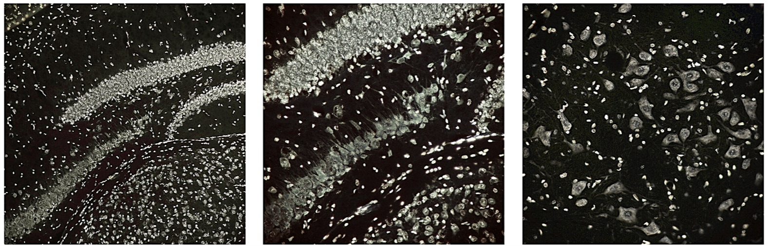
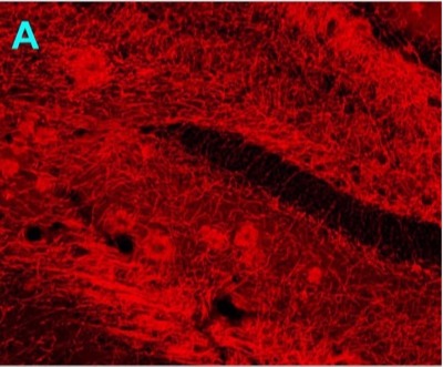
Ethidium Bromide-Red 

This tracer stains the cytoplasm and nuclei of neurons a fluorescent red color under green light excitation. The stain is of high contrast, resolution and brightness and is also very resistant to fading. Although most visible under green light excitation, this fluorochrome is moderately visible under blue light excitation and faintly visible under UV excitation.

Recently Developed

Neutral Red-Red

This fluorochrome stains the cytoplasm and nuclei of neurons a fluorescent red color under green light excitation. The stain is of relatively high contrast, resolution and is moderately resistant to fading. The fluorochrome shows minimal bleed-through when excited by shorter wavelengths such as blue or UV light.

Recently Developed

Acridine Orange-Green

This fluorochrome stains the cytoplasm and nuclei of neurons a fluorescent green color under blue light excitation. The stain is of high contrast and resolution. The fluorochrome exhibits minimal bleed through when excited by longer or shorter wavelengths and is moderately resistant to fading.

Recently Developed

Hydroxystilbamidine-Silver

This fluorochrome results in the cytoplasm and nuclei of neurons staining a fluorescent near white color under ultraviolet light excitation. The stain is of very high contrast, brightness and resolution. Also, it is not visible under longer wavelengths, such as blue or green light excitation. It is also highly resistant to fading.

Recently Developed

Euro-Glo ®

This tracer will label both myelinated fibers and amyloid plaques. Although it has been available for over a year, it has not been extensively used, possibly because it differs from most of the other tracers that target A-beta aggregates. By contrast, Euro-Glo is thought to label glycolipids, such as gangliosides, resulting in a different labeling pattern. The relatively long staining times (5-7 days) required may be less convenient than more rapid stains such as Amylo-Glo. This compound represents the only histochemical stain for use labeling brain tissue sections whose fluorescence is based on the presence of a rare earth metal (Europium) as opposed to an organic fluorophore.

Recently Developed

HQ-O

A recently published study characterized this tracer’s ability to form a fluorescent precipitate with zinc and in doing so giving a high contrast and resolution staining of amyloid plaques. The staining time is of intermediate length, typically between 1 and 24 hours, depending on the temperature of the stain and the nature of prior tissue processing.

Upcoming

Amylo-Glo ® II

This tracer, like the original Amylo-Glo, will stain both extracellular amyloid plaques and intracellular neurofibulary tangles in both AD human autopsy and transgenic mouse brain tissue. The primary difference between Amylo-Glo and Amylo-Glo II is that the former is available as a 10X solution containing NaOH (final pH = 10.5), while Amylo-Glo II is offered as a powder that can be reconstituted in a vehicle of 50% ethanol. Although both tracers exhibit essentially the same labeling pattern, the Amylo-Glo II is more stable in solution and results in labeling of noticeably higher contrast, as illustrated to the left.